
What Is Ultrasonography?
Ultrasonography is one of the most widely used imaging methods in modern healthcare. It works through sound waves that create detailed images of internal organs, soft tissues, and blood vessels. This method does not use radiation, which makes it safe even for pregnant women and children. It provides real-time visuals that help doctors observe organ structure and detect possible abnormalities. The process is simple and completely painless. A clear gel is applied to the skin before scanning to help sound waves pass smoothly through the body. Because of its accuracy and safety, ultrasonography is often used for early diagnosis and medical monitoring in many fields, including cardiology, gynecology, and general surgery.
Ultrasonography in Turkey
Ultrasonography in Turkey is an essential part of diagnostic medicine. Modern hospitals and imaging centers across the country use advanced ultrasound systems for both general and specialized scans. Radiologists and trained technicians perform these procedures carefully to ensure precise imaging results. Turkey’s medical facilities are known for offering reliable, patient-centered diagnostic care. Many people undergo routine check-ups, abdominal or pelvic scans, and pregnancy monitoring through ultrasound examinations. In addition to local patients, international visitors also trust Turkey’s healthcare system for its modern technology and professional medical approach.
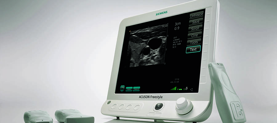
How Does Ultrasonography Work?
The principle of ultrasonography is based on high-frequency sound waves that move through the body and reflect back from tissues and organs. A device called a transducer both sends and receives these waves, which are then transformed into visual images by computer software. The quality of these images depends on the density of the tissues and the type of probe used. Since the method does not involve radiation, it can be repeated when necessary for follow-up assessments. The scanning process is quick, lasting only a few minutes depending on the area examined. Patients usually do not need any special preparation except for certain abdominal scans that may require fasting or a full bladder.
Types of Ultrasound Scans
Ultrasound imaging includes several types, each designed for specific medical purposes. Abdominal ultrasound helps examine the liver, pancreas, gallbladder, and kidneys. Pelvic ultrasound is often used to check the uterus, ovaries, and bladder. Obstetric ultrasound monitors fetal growth and development during pregnancy. Doppler ultrasound measures blood flow, helping detect vascular blockages or circulation problems. In addition, thyroid and breast ultrasounds provide detailed images for detecting nodules or cysts. More advanced types, such as 3D and 4D ultrasounds, offer detailed and dynamic visuals that support both medical evaluation and patient reassurance. Each form of ultrasonography contributes to accurate diagnosis and effective treatment planning.
When Is Ultrasonography Used?
This imaging method is applied in many areas of medicine to evaluate organs, detect diseases, and monitor treatments. It helps assess conditions in the abdomen, pelvis, thyroid, and heart, as well as blood flow in veins and arteries. Obstetric ultrasound is essential for observing pregnancy development and the baby’s health. Musculoskeletal ultrasound is also useful for diagnosing joint or tendon problems. In emergency cases, it provides quick insights without exposing patients to radiation. Because it is versatile, ultrasonography is often chosen for both preventive check-ups and ongoing medical evaluations, allowing doctors to make accurate clinical decisions based on real-time images.
Benefits of Ultrasonography
The biggest advantage of ultrasonography is that it is completely non-invasive and painless. It does not require any injections or contrast agents, and it avoids radiation exposure. The procedure can be repeated safely whenever follow-up imaging is needed. Real-time images make it easier to guide biopsies or medical procedures with precision. It also offers immediate results, which can be especially important in urgent medical situations. The clarity of the images helps detect changes in tissues or organs at an early stage. Additionally, it supports doctors in tracking the progress of ongoing treatments and ensuring accurate diagnosis with minimal discomfort for the patient.
Is Ultrasonography Safe?
Ultrasonography is considered one of the safest diagnostic tools available today. It uses high-frequency sound waves, not ionizing radiation, which means there is no known risk of tissue damage. For this reason, it is widely used in prenatal care, as it poses no harm to the mother or the developing baby. The procedure does not cause pain, allergic reactions, or long-term side effects. Since it is performed externally, it does not require recovery time. Even for patients with chronic conditions, ultrasonography remains a safe and reliable imaging choice that can be repeated whenever necessary under medical supervision.
What to Expect During an Ultrasound Exam
An ultrasound exam is a simple and comfortable process that usually takes between 10 and 30 minutes. The patient is asked to lie down while a clear gel is applied to the skin to help sound waves travel effectively. The technician or doctor gently moves a transducer over the area being examined. This device sends sound waves into the body and instantly displays the returning echoes as images on a screen. Patients may feel mild pressure but no pain throughout the procedure. Once the scan is complete, the gel is wiped off and normal daily activities can be resumed immediately. The results are typically reviewed by a radiologist and then shared with the attending physician for evaluation.
How to Prepare for an Ultrasound
Preparation for an ultrasound depends on the type of examination being performed. In most cases, it requires little to no special preparation. For abdominal scans, patients may be asked to fast for several hours to reduce gas in the intestines, which can interfere with image clarity. In pelvic or obstetric ultrasounds, drinking plenty of water before the exam helps fill the bladder and improves image visibility. Comfortable clothing is recommended, as some areas may need to be exposed during the procedure. Jewelry and metallic accessories should be removed to avoid interference with the equipment. Following the preparation instructions ensures more accurate and reliable imaging results.
Limitations of Ultrasonography
Although ultrasonography is a valuable imaging method, it does have some limitations. Sound waves cannot pass through bone or air-filled spaces effectively, which makes it difficult to examine certain structures like the lungs or areas hidden behind bone. In obese patients, excess tissue may reduce image quality. Some abnormalities may require further evaluation with MRI or CT scans for more detailed results. The accuracy of the examination can also depend on the experience of the radiologist and the type of device used. Despite these limitations, ultrasonography remains one of the most accessible, safe, and effective tools for diagnostic imaging in clinical practice.
Ultrasonography in Turkey Cost 2025
The average cost of ultrasonography in Turkey in 2025 varies depending on the type of scan and the region of the body being examined. Basic ultrasounds such as abdominal or thyroid scans generally range between 40 and 80 USD, while more specialized procedures like Doppler or pregnancy ultrasounds can cost between 70 and 150 USD. Prices may change based on the city, the complexity of the procedure, and whether the scan is part of a larger diagnostic evaluation. Many medical centers provide detailed imaging with modern equipment and trained specialists, ensuring accurate results at reasonable rates compared to global averages.
Is Ultrasound the Same as Ultrasonography?
The terms “ultrasound” and “ultrasonography” are often used interchangeably, but there is a slight difference in meaning. “Ultrasound” refers to the sound waves themselves or the imaging process in general, while “ultrasonography” is the complete diagnostic procedure that uses these waves to produce images of internal organs and tissues. In clinical settings, both terms describe the same method used for medical imaging. The distinction is mainly technical, and in everyday medical communication, either term can be used without confusion. Both describe a safe, non-invasive approach that helps physicians examine the body in real time and detect a wide range of health conditions.
Does Ultrasonography Use Radiation?
Ultrasonography does not use radiation. Instead, it relies on high-frequency sound waves to create images of internal organs and tissues. These sound waves are harmless and cannot cause any cellular damage, making the method completely safe for patients of all ages. Because no ionizing radiation is involved, ultrasonography is also suitable for pregnant women and newborns. The procedure can be repeated when necessary without risk to the body. For this reason, it has become one of the most trusted and frequently used diagnostic tools in modern medicine, preferred for both routine and advanced medical evaluations.
Can You Eat Before an Abdominal Ultrasound?
For most abdominal ultrasound examinations, eating or drinking before the procedure is not recommended. Food and gas in the digestive tract can affect image quality and make it harder to visualize organs like the liver, gallbladder, and pancreas. Patients are usually asked to avoid eating for 6 to 8 hours before the exam to ensure clear results. However, small sips of water may be allowed if necessary. In some cases, instructions can vary depending on the area to be scanned. Following the preparation guidelines provided by the healthcare professional helps achieve the most accurate and reliable diagnostic outcome.
How Long Does an Ultrasound Scan Take?
An ultrasound scan is generally quick, taking between 10 and 30 minutes depending on the type of examination. Shorter scans, such as thyroid or breast imaging, may finish in just a few minutes, while detailed abdominal or Doppler studies can take longer. The process begins with applying a gel to the skin, which helps the transducer move smoothly and improves image quality. The technician carefully moves the device over the body, capturing real-time images. After the scan, the gel is wiped off, and the patient can return to normal activities immediately. The results are usually analyzed shortly after the procedure for review by the physician.
Is Ultrasonography Painful?
Ultrasonography is a completely painless procedure. Most patients only feel light pressure when the transducer moves over the skin. The gel used during the scan may feel slightly cool, but it does not cause discomfort. Because there are no injections, cuts, or radiation involved, the process is simple and relaxing. In rare cases, mild pressure might cause temporary sensitivity if the examined area is already tender, but it disappears quickly after the scan. Patients can resume their daily routines immediately after the procedure, making ultrasonography one of the most comfortable and stress-free imaging techniques in medical practice.
