

What Is Mammography?
Mammography is a specialized imaging technique used to examine breast tissue for early signs of disease, particularly breast cancer. It allows doctors to detect small lumps or changes that cannot be felt during a physical examination. The method uses low-dose X-rays to capture detailed images of the breast, helping identify abnormalities such as calcifications, cysts, or tumors. Early detection through mammography greatly increases the chances of successful treatment. The procedure is recommended as part of regular health screenings for women, especially those over the age of 40 or with a family history of breast cancer.
Mammography in Turkey
Mammography in Turkey is a routine part of women’s health check-ups and preventive care. Many hospitals and diagnostic centers use modern digital mammography systems that provide high-resolution images with minimal radiation exposure. Radiologists trained in breast imaging carefully evaluate each scan for any sign of irregularity. The procedure is performed in a private and comfortable environment to ensure the patient feels at ease. With growing awareness of early cancer detection, mammography has become an essential and accessible tool for maintaining women’s health throughout the country.
How Does a Mammogram Work?
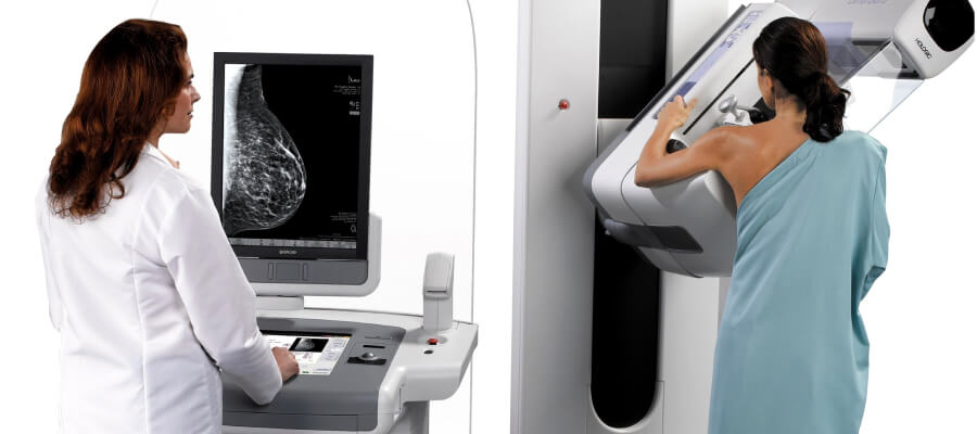
A mammogram works by using low-dose X-ray technology to create images of breast tissue. During the procedure, each breast is gently positioned and compressed between two flat plates for a few seconds. This compression spreads the tissue evenly, allowing clearer and more detailed images while reducing the radiation dose. The process might cause slight pressure but does not cause lasting discomfort. Once the images are taken, they are reviewed by a radiologist who looks for any unusual patterns or structures. The procedure usually takes less than 20 minutes, and results are analyzed soon after the examination.
Types of Mammograms: 2D vs 3D
There are two main types of mammograms used in breast imaging: 2D and 3D mammography. Traditional 2D mammography captures two flat images of each breast, one from the top and one from the side. While effective, it can sometimes make it difficult to distinguish overlapping tissues. 3D mammography, also known as digital breast tomosynthesis, creates multiple thin-layer images that provide a more detailed and accurate view. This advanced method improves cancer detection, especially in women with dense breast tissue. Both techniques are safe and valuable, but 3D imaging offers clearer visualization for more confident diagnosis.
When Should You Get a Mammogram?
A mammogram should generally be done once a year or every two years, depending on a woman’s age, risk factors, and medical history. Most health experts recommend that women begin regular screenings at the age of 40, but those with a family history of breast cancer may start earlier. The ideal timing also depends on hormonal changes, such as menopause or hormone therapy. Scheduling the test a week after the menstrual period often helps minimize breast tenderness during the procedure. Regular mammograms allow for early detection of any changes, improving the chances of successful and less invasive treatment.
Benefits of Mammography for Early Detection
The main benefit of mammography lies in its ability to detect breast cancer before symptoms appear. Early detection increases treatment options and significantly improves recovery rates. Mammography can reveal small tumors, microcalcifications, or structural changes that cannot be felt by touch. By identifying these signs early, doctors can intervene before the disease progresses. Regular screenings also provide valuable baseline comparisons, making it easier to notice subtle differences over time. This method not only saves lives but also supports better long-term outcomes through timely and accurate diagnosis.
Is Mammography Safe?
Mammography is a safe and reliable imaging method that uses a very low dose of X-rays. The level of radiation exposure is minimal and well within international safety standards. The benefits of early detection far outweigh any potential risks associated with the procedure. Modern digital mammography systems are designed to reduce radiation further while providing high-quality images. Some women may feel brief pressure or mild discomfort during the scan, but it quickly subsides once the images are taken. The procedure is non-invasive, and patients can safely resume normal activities immediately after the examination.
What to Expect During a Mammogram
During a mammogram, each breast is placed on a flat surface and gently compressed with a transparent plate. This compression helps spread the tissue evenly to produce clearer images. The procedure is quick, usually lasting around 15 to 20 minutes. A trained technician ensures the process is as comfortable as possible. Some women may feel mild pressure, but it does not cause pain or harm. Once the images are captured, they are reviewed by a radiologist who examines them for any irregularities. Patients can expect to receive their results shortly afterward, along with a clear explanation from their healthcare provider.
How to Prepare for a Mammogram
Preparing for a mammogram involves a few simple steps to ensure accurate and comfortable imaging. It is best to schedule the exam for a time when the breasts are least sensitive, usually a week after the menstrual period. On the day of the scan, patients should avoid using deodorants, powders, lotions, or perfumes under the arms or on the chest, as these can appear as white spots on the X-ray image. Comfortable two-piece clothing is recommended so the upper body can be easily exposed. Any previous mammogram images should be brought to the appointment for comparison. Informing the technician about any breast symptoms, surgeries, or implants helps ensure proper evaluation and accurate results.
Understanding Mammogram Results
Mammogram results are analyzed by a radiologist who carefully examines the breast tissue for any changes or unusual patterns. The findings are typically categorized using the BI-RADS (Breast Imaging Reporting and Data System) scale, which ranges from normal results to findings that may need further evaluation. Most mammograms show normal tissue or benign conditions like cysts or calcifications. If something unusual is detected, additional imaging or tests such as ultrasound or biopsy may be recommended. A clear and detailed report is then shared with the doctor, who explains the results to the patient. Understanding these findings helps patients stay informed and confident about their breast health.
Mammography in Turkey Cost 2026
In 2026, the average cost of a mammography examination in Turkey ranges between 40 and 100 USD, depending on the city, clinic, and technology used. Digital and 3D mammography systems may cost slightly more but offer enhanced image quality and better detection accuracy. Prices may vary if additional diagnostic views or follow-up imaging are needed. Many diagnostic centers provide professional breast imaging with advanced equipment and trained radiologists. Regular screenings remain one of the most effective ways to detect breast cancer early, and mammography in Turkey continues to offer a reliable and affordable option for maintaining long-term women’s health.
Does a Mammogram Hurt?
A mammogram may cause brief discomfort, but it should not be painful. During the procedure, each breast is gently compressed between two plates for a few seconds to create clear and detailed images. This compression can feel slightly uncomfortable, especially for those with sensitive breasts, but it lasts only a short time. Taking slow breaths and relaxing the muscles can help reduce tension during the scan. Scheduling the exam a week after the menstrual period also helps minimize sensitivity. The momentary pressure is a small inconvenience compared to the significant benefits of early detection and breast health monitoring.
How Often Should You Get a Mammogram?
The recommended frequency for mammograms depends on age, health history, and risk factors. Most experts advise women to begin regular screenings at the age of 40 and continue every one to two years. Those with a higher risk, such as a family history of breast cancer or genetic predisposition, may start earlier or undergo more frequent check-ups. Maintaining a consistent screening schedule allows doctors to identify subtle changes over time and detect potential issues at the earliest stage. Regular mammograms are a vital part of preventive healthcare for women in all age groups.
Can I Wear Deodorant Before a Mammogram?
Wearing deodorant, lotion, perfume, or powder before a mammogram is not recommended. These products can contain metallic or powdery substances that may appear as white spots on X-ray images, potentially leading to confusion or inaccurate readings. It is best to clean the underarm area before the appointment and avoid applying anything to the chest or armpits. If deodorant is accidentally used, the technician can provide wipes to remove it before the scan. Following these simple steps ensures that the images are clear and the evaluation is precise.
Are Mammograms Accurate for Dense Breasts?
Mammograms can detect many abnormalities even in women with dense breast tissue, but dense tissue can sometimes make it harder to identify small lesions. Dense breasts contain more glandular and connective tissue, which can appear white on a mammogram, similar to tumors. In such cases, doctors may recommend additional imaging tests such as breast ultrasound or MRI for a more detailed view. Combining these methods improves accuracy and ensures thorough evaluation. Regular screenings and open communication with healthcare providers help maintain effective monitoring, regardless of breast density.
What Happens If My Mammogram Is Abnormal?
An abnormal mammogram does not automatically mean cancer. It indicates that something unusual was seen, and further evaluation is needed. The radiologist may suggest additional imaging, such as magnified mammography, ultrasound, or biopsy, to better understand the findings. These follow-up tests help determine whether the changes are benign or require treatment. Most abnormalities turn out to be harmless conditions like cysts or calcifications. After further analysis, the doctor explains the results clearly and discusses the next steps. Early and detailed investigation ensures the best possible outcome for breast health.
