
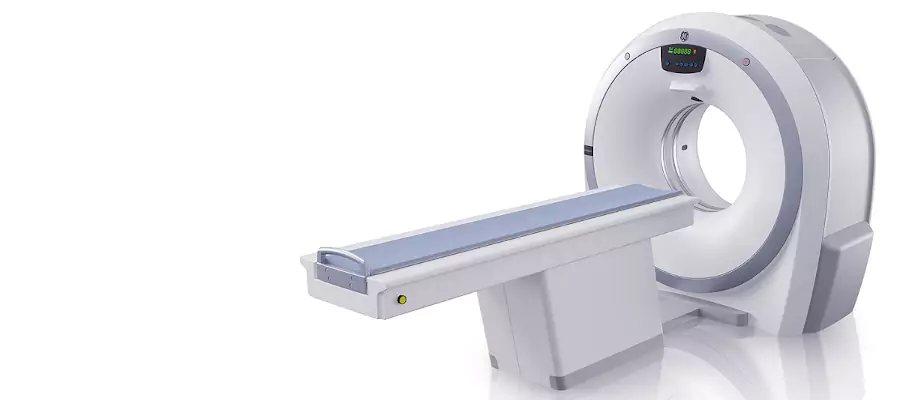
What Is Computed Tomography (CT Scan)?
A computed tomography (CT) scan is an advanced medical imaging test that combines multiple X-ray images to create detailed cross-sectional views of the body. It allows doctors to examine bones, organs, blood vessels, and soft tissues with high precision. The images are processed by a computer to form 3D visuals, helping detect tumors, fractures, infections, or internal bleeding. CT scans are often used for quick diagnosis in emergency cases or to guide surgical and treatment planning. The procedure is painless, fast, and widely used across different medical specialties to provide accurate and comprehensive information about a patient’s condition.
Computed Tomography (CT Scan) in Turkey
CT scanning is a commonly used diagnostic method in Turkey, available in hospitals and imaging centers equipped with modern technology. Radiologists perform and interpret these scans with expertise, ensuring reliable and high-quality results. The procedure plays an important role in diagnosing a wide range of conditions, from head injuries and chest diseases to abdominal or bone problems. In Turkey, CT services are performed under safe and standardized protocols that minimize radiation exposure. The process is completed within minutes, making it ideal for both emergency situations and planned evaluations requiring clear internal imaging.
How Does a CT Scan Work?
A CT scan works by sending a series of X-ray beams through the body from different angles. As the scanner rotates around the patient, detectors capture the data and send it to a computer, which reconstructs detailed cross-sectional images. These slices can be viewed individually or combined into a 3D model, allowing doctors to see internal structures clearly. Depending on the type of scan, a contrast dye may be used to highlight specific organs or blood vessels. The entire process is fast and non-invasive, providing accurate insights that help guide diagnosis and medical treatment effectively.
CT Scan vs MRI: What’s the Difference?
Although both CT and MRI scans produce detailed internal images, they use different technologies. A CT scan relies on X-rays and is particularly effective for viewing bones, detecting fractures, or identifying bleeding and lung conditions. An MRI uses magnetic fields and radio waves, making it better suited for examining soft tissues such as the brain, muscles, and ligaments. CT scans are faster and often preferred in emergency cases, while MRIs provide more detail for neurological or musculoskeletal evaluations. Both methods complement each other, allowing doctors to choose the most suitable technique depending on the medical need.
When Do You Need a CT Scan?
A CT scan is performed when a detailed internal image is needed to diagnose or monitor a medical condition. It helps detect fractures, infections, blood clots, tumors, and internal bleeding. Doctors often request it after an accident or injury to check for damage to the brain, chest, or abdomen. It is also used to plan surgeries, evaluate treatment results, or guide biopsies. In some cases, a CT scan can reveal conditions that are not visible on standard X-rays. Because of its accuracy and speed, it plays an essential role in both emergency care and routine diagnostic assessments.
Common Types of CT Scans
CT imaging can be performed on different parts of the body depending on the medical concern. A head CT scan detects strokes, tumors, and brain injuries. A chest CT examines the lungs, heart, and blood vessels. An abdominal and pelvic CT helps identify organ problems such as kidney stones, appendicitis, or liver disease. A spinal CT is useful for assessing disc herniation and vertebral fractures. Some procedures use contrast-enhanced CT scans to highlight blood flow and organ structure more clearly. Each type provides specific information that supports accurate diagnosis and targeted medical treatment.
Is a CT Scan Safe? (Radiation Risks Explained)
A CT scan involves a small amount of ionizing radiation, but the exposure level is carefully controlled and considered safe for most patients. The benefits of accurate diagnosis usually outweigh any potential risk. Modern CT scanners use advanced technology to minimize radiation doses while maintaining image quality. Pregnant women should inform their doctor before the test so that alternative imaging methods can be considered. In rare cases, patients may experience mild reactions to contrast dye, such as warmth or metallic taste, but these effects disappear quickly. Overall, CT scanning remains a safe and reliable tool when performed under proper medical supervision.
What to Expect During a CT Scan Procedure
During a CT scan, the patient lies on a motorized table that moves slowly through a circular scanner. Depending on the test type, a contrast dye may be injected into a vein to help highlight internal structures. The scan itself is quick, typically lasting 5 to 15 minutes. The patient must remain still to ensure clear images, though the procedure is completely painless. The machine may produce soft whirring sounds while capturing images from different angles. Once the scan is complete, patients can resume their daily activities right away. The results are reviewed by a radiologist and then shared with the doctor for detailed evaluation.
How to Prepare for a CT Scan
Preparation for a CT scan depends on the area being examined and whether contrast material will be used. Patients are usually advised to avoid eating or drinking for four to six hours before the test if a contrast dye is required. Comfortable clothing without zippers or metal objects is recommended, and all jewelry or accessories should be removed before the scan. People with allergies, kidney problems, or previous reactions to contrast material should inform their doctor beforehand. Pregnant women should also notify the medical team as a precaution. Following these simple instructions ensures that the images are clear and the procedure is completed safely and efficiently.
How Long Does a CT Scan Take?
A CT scan is a quick and efficient imaging procedure, typically lasting between 5 and 30 minutes depending on the type of study. The actual scanning process often takes just a few minutes, but additional time may be needed for preparation or the administration of contrast dye. Patients lie on a table that slowly moves through the scanner while X-ray images are captured from multiple angles. It is important to stay still to prevent blurring. After the scan, the patient can return to normal activities right away. The images are then analyzed by a radiologist and reviewed by the doctor for diagnosis.
Computed Tomography in Turkey Cost 2026
In 2026, the average cost of a CT scan in Turkey ranges between 70 and 200 USD, depending on the type of scan, the use of contrast material, and the region of the body being examined. Advanced imaging centers across the country use modern CT technology that ensures high-resolution results with minimal radiation exposure. Head, chest, abdominal, and whole-body CT scans are commonly performed as part of diagnostic evaluations. Prices may vary slightly between public hospitals and private imaging centers, but all follow strict safety and quality standards. The combination of accuracy, accessibility, and reasonable pricing makes CT scanning an essential diagnostic tool in Turkey’s healthcare system.
Do I Need Contrast Dye for a CT Scan?
Not every CT scan requires contrast dye, but it may be used when doctors need to highlight specific organs or blood vessels. The contrast material helps make structures such as the intestines, brain, or kidneys appear more clearly on the images. It can be given orally, intravenously, or both, depending on the purpose of the scan. The substance used is generally safe, and any mild sensations like warmth or a metallic taste disappear quickly. People with kidney problems or allergies should inform their doctor beforehand. The decision to use contrast is made based on medical necessity to ensure accurate diagnostic results.
Can I Eat or Drink Before a CT Scan?
Eating or drinking before a CT scan depends on whether contrast dye will be used. For scans without contrast, normal eating and drinking are usually allowed. However, if a contrast-enhanced scan is planned, fasting for four to six hours before the procedure is often recommended to prevent nausea and ensure clear imaging. Drinking water is typically permitted and even encouraged. Patients should follow the specific instructions provided before the appointment. Clear preparation helps produce high-quality images and minimizes the need for repeat scanning.
Is a CT Scan Painful?
A CT scan is a completely painless and non-invasive procedure. Patients may feel slight discomfort when lying still on the table or if a contrast dye is injected, but the sensation is brief and mild. The scan itself does not cause any pain, and most people find it easy to tolerate. The equipment makes soft rotating sounds while capturing images, which is normal. The process is quick, and patients can resume regular activities immediately afterward. Overall, CT scanning is known for being safe, comfortable, and efficient for diagnosing a wide range of conditions.
How Soon Do CT Scan Results Come Back?
CT scan results are usually available within 24 to 48 hours, depending on the healthcare facility and the complexity of the images. In urgent cases, preliminary results may be reviewed immediately. A radiologist carefully examines the images, looking for any signs of abnormality such as inflammation, bleeding, or growths. The findings are then compiled into a detailed report and shared with the referring doctor, who discusses the results with the patient. This step-by-step process ensures that the diagnosis is accurate and that appropriate treatment decisions can be made promptly.
Can a CT Scan Detect Cancer?
A CT scan can help detect cancer by showing detailed images of tumors, abnormal tissue growth, or organ changes. It is often used to locate the size and position of a tumor, determine whether it has spread, and evaluate how well treatment is working. CT scans are commonly performed for cancers of the lungs, liver, kidneys, pancreas, and abdomen. While the scan alone may not confirm a diagnosis, it provides essential information that guides further testing, such as biopsy or MRI. Its precision and clarity make it a vital tool in cancer detection and management.
