
What Is a DEXA Scan?
A DEXA scan, also known as Dual-Energy X-ray Absorptiometry, is a medical imaging test used to measure bone density. It helps determine the strength and mineral content of bones, allowing early detection of osteoporosis or other bone-related conditions. The procedure is quick, non-invasive, and painless. It uses two low-dose X-ray beams at different energy levels to capture images of bones, usually in the lower spine and hips. By analyzing how much of each beam passes through the bone, doctors can calculate bone density accurately. The results are essential for assessing fracture risk and monitoring bone health over time.
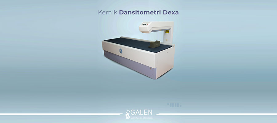
DEXA Scan in Turkey
DEXA scan in Turkey are widely performed in hospitals and radiology centers as part of preventive health care and osteoporosis management. The procedure is carried out by trained radiology technicians and interpreted by experienced physicians. DEXA technology is especially valuable for postmenopausal women, elderly individuals, and patients with hormonal or metabolic conditions affecting bone strength. Modern facilities in Turkey offer precise bone density measurements with comfortable and fast scanning procedures. Routine DEXA scans are often recommended as part of annual health check-ups to evaluate bone health and prevent long-term complications.
What Does a DEXA Scan Measure?
A DEXA scan measures bone mineral density (BMD), which reflects the amount of calcium and other minerals in bones. The results are given as T-scores and Z-scores that compare a person’s bone strength to healthy reference values. Low T-scores indicate reduced bone mass and a higher risk of fractures, which may signal osteopenia or osteoporosis. In addition to bone density, modern DEXA systems can also measure body composition, including fat and muscle distribution. This information helps doctors create personalized treatment plans, track changes in bone health, and evaluate how lifestyle or medications affect bone strength.
DEXA Scan vs Bone Scan: What’s the Difference?
While both DEXA scans and bone scans assess bone health, they serve different purposes. A DEXA scan measures bone density to evaluate strength and detect conditions like osteoporosis. It uses low-dose X-rays and provides numerical results that reflect bone mineral levels. A bone scan, on the other hand, detects abnormalities such as fractures, infections, or tumors within the bone using small amounts of radioactive material. In summary, DEXA focuses on bone quality and density, while a bone scan identifies changes in bone metabolism or structure. Both tests complement each other in providing a complete view of bone health.
Who Should Get a DEXA Scan?
A DEXA scan is recommended for individuals who may be at risk of weakened bones or osteoporosis. Women over the age of 50 and men over 65 are typically advised to have regular bone density checks. It is also beneficial for people with a family history of fractures, hormonal disorders, or medical conditions affecting calcium absorption. Those taking long-term corticosteroid medication or who have experienced unexplained fractures may also need evaluation. Postmenopausal women and individuals with low body weight or vitamin D deficiency benefit most from early testing. Regular DEXA scans help track bone strength and support timely preventive care before major complications occur.
Benefits of a DEXA Scan
The greatest advantage of a DEXA scan is its ability to detect bone loss before it leads to fractures. It provides a precise measurement of bone mineral density, allowing doctors to assess the risk of osteoporosis and monitor treatment progress. The test is fast, painless, and involves very low radiation exposure. By identifying even small decreases in bone density, it enables early intervention through lifestyle changes or medication. The scan can also assess body composition, showing fat and muscle ratios that help design balanced health plans. Regular DEXA evaluations play an important role in maintaining bone health and preventing age-related bone conditions.
Is a DEXA Scan Safe?
A DEXA scan is considered one of the safest imaging procedures in medicine. It uses a minimal dose of X-rays, far lower than that of a standard chest X-ray. The test is non-invasive, does not require injections, and causes no discomfort. There are no known long-term side effects, and the radiation exposure is negligible, even for repeated scans. However, pregnant women should inform their doctor before the test as a precaution. The procedure is suitable for almost everyone, providing accurate and reliable bone health information without any health risks.
What to Expect During a DEXA Scan
During a DEXA scan, the patient lies comfortably on a padded table while a scanning arm passes slowly over the body. The test usually focuses on areas like the hip and lower spine, where bone loss commonly occurs. No special preparation is required, although removing metal objects such as belts or jewelry helps prevent image interference. The procedure takes about 10 to 20 minutes and is completely painless. Once the scan is complete, results are reviewed by a radiologist who calculates bone density scores. These findings help the doctor determine bone strength, assess fracture risk, and plan any necessary treatment or lifestyle adjustments.
How to Prepare for a DEXA Scan
Preparation for a DEXA scan is simple and requires little effort. Patients can eat and drink normally before the test, but calcium supplements should be avoided for at least 24 hours prior to the appointment, as they may affect results. Comfortable clothing without metal buttons, zippers, or belts is recommended, since metal can interfere with X-ray imaging. Jewelry and accessories should be removed before the procedure. If the patient has recently undergone a barium study or received a contrast injection, it is best to wait several days before having the DEXA scan. Informing the technician about medical history, medications, or possible pregnancy ensures a safe and accurate assessment.
How to Read DEXA Scan Results (T-score & Z-score Explained)
DEXA scan results are presented in two key values: the T-score and the Z-score. The T-score compares a patient’s bone density to that of a healthy young adult. A score above -1 is considered normal, between -1 and -2.5 indicates osteopenia (mild bone loss), and below -2.5 suggests osteoporosis. The Z-score, on the other hand, compares bone density to people of the same age, sex, and body size. A low Z-score may indicate bone loss caused by an underlying condition rather than age alone. These measurements help doctors identify bone health risks and recommend suitable lifestyle changes or treatments to protect and strengthen bone structure.
DEXA Scan in Turkey Cost 2025
The average cost of a DEXA scan in Turkey in 2025 ranges between 30 and 70 USD, depending on the region and type of medical center. Prices can vary slightly based on whether the scan includes additional body composition analysis or multiple body areas. Hospitals and radiology clinics across Turkey perform DEXA scans using advanced imaging technology that ensures accurate and detailed results. The procedure is quick, affordable, and widely available, making it an important part of preventive healthcare for both local and international patients. Regular bone density evaluations through DEXA scanning help maintain long-term bone strength and prevent future fractures.
At What Age Should You Get a DEXA Scan?
A DEXA scan is typically recommended for women over the age of 50 and men over 65, as bone density naturally decreases with age. However, younger individuals may also need testing if they have certain risk factors such as hormonal imbalances, long-term use of corticosteroids, a family history of osteoporosis, or frequent fractures. Postmenopausal women and people with low body weight or chronic medical conditions affecting bone strength benefit from early evaluation. Having a baseline DEXA scan allows doctors to monitor bone health over time and take preventive measures before serious bone loss occurs.
How Long Does a DEXA Scan Take?
A DEXA scan is one of the quickest imaging procedures in modern medicine, usually taking between 10 and 20 minutes. The duration may vary slightly depending on which body areas are scanned, most commonly the hip and spine. The process involves lying still on a padded table while a scanner passes over the body. The procedure is painless and does not require any recovery time. After the scan, the images are analyzed by a radiologist, and results are usually available shortly afterward. Patients can return to their normal daily activities immediately following the exam.
Can I Eat Before a DEXA Scan?
Eating before a DEXA scan is generally allowed, as food does not interfere with the imaging process. However, calcium supplements should be avoided for at least 24 hours before the test because they can temporarily affect bone density readings. Drinking water is encouraged, but it is best to avoid wearing clothing with metal parts that could interfere with the X-ray beam. Patients should also inform the technician if they have recently undergone other imaging tests that used contrast materials. Following these simple guidelines ensures a smooth and accurate examination.
Is a DEXA Scan Covered by Insurance?
Many insurance plans cover DEXA scans, especially when the test is medically necessary for evaluating bone health or diagnosing osteoporosis. Coverage may depend on factors such as age, gender, risk level, and doctor’s recommendation. Some plans allow routine screening for women over 50 or patients with known bone loss conditions. It is always best to check the policy details or consult the insurance provider beforehand to understand any requirements or limitations. Even without full coverage, the scan remains an affordable and valuable tool for preventive health care and long-term bone protection.
How Often Should You Have a DEXA Scan?
The frequency of DEXA scans depends on bone density results and personal risk factors. For most adults with normal bone health, a scan every two to three years is sufficient. Those with osteopenia or osteoporosis may need more frequent evaluations, usually once a year, to monitor changes and adjust treatment if necessary. Individuals taking medication for bone loss should also have regular scans to measure improvement. Consistent monitoring helps detect even small changes in bone strength, allowing early intervention and reducing the risk of fractures or advanced bone disease.
